
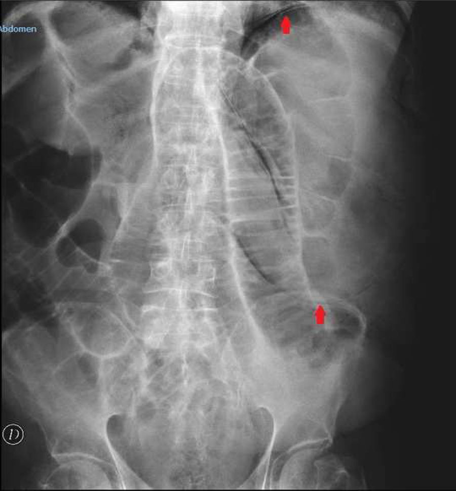
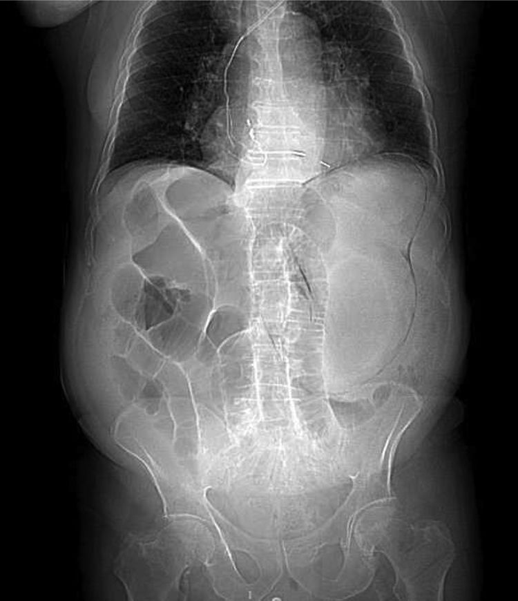
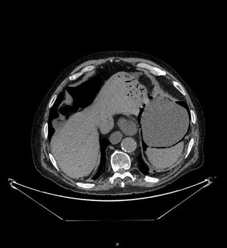
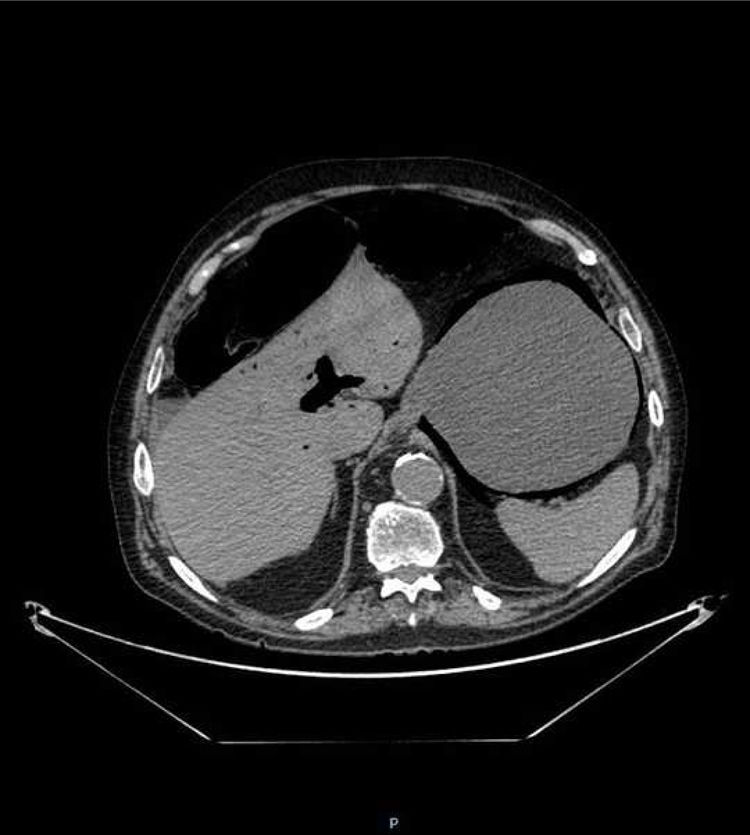
An 82-year-old man presented with bloating and generalized abdominal pain, accompanied by vomiting (dark in color, with an aspect of retention vomiting) and diarrheic stools. A plain abdominal x-ray (Fig. 1) showed air in the gastric wall and bowel segment dilation. An abdominal computed tomography (CT) scan was ordered (Fig. 2) that revealed intramural gas associated with gastric dilation, pneumoperitoneum (Fig. 3), and gas in the portal vein (Fig. 4). An exploratory laparotomy was performed, finding a distended stomach with gas bubbles in its serosa and no signs of transmural necrosis or perforation. Intraoperative gastroscopy identified ulcerated-necrotic mucosa with no spontaneous bleeding on the posterior surface, body, and greater curvature. Given those findings, no further surgical act was carried out. Lactobacillus jensenii was isolated in blood cultures and treated with meropenem + linezolid. The patient progressed favorably, with improvement in the control abdominal CT scan, and was released from the hospital. Emphysematous gastritis is a rare pathology produced by the translocation of gas-producing microorganisms in the walls of the stomach. The causal agent cannot be isolated in up to 42.4%1 of cases and there is a 60% mortality rate.2 CT is the diagnostic study of choice and is essential for making early diagnosis and implementing the vital support treatment with broad-spectrum antibiotics. Surgery is only required if there is no response to conservative treatment or in cases of severe sepsis or gastric perforation.3
 and small bowel segment dilation.")


Axial view of the abdominal CT scan with iv contrast medium showing abundant quantities of intraparietal gas in the stomach, small bowel segment dilation with a maximum caliber of 4.3 cm, and a pneumoperitoneum of moderate quantity adjacent to the gastric body and anterior abdominal wall.
.")
The authors declare that no experiments were conducted on animals or humans for the present research and that they followed the protocols of their work center on the publication of patient data, preserving patient anonymity at all times, guaranteeing that the article contains to personal information that could identify patients. The authors received informed consent from the patient described in the article and that document is in the possession of the corresponding author. Likewise, the study meets the current standards in bioethical research, adhering to the protocols and in accordance with the patient and his family.
Financial disclosureNo financial support was received in relation to this article.
Conflicts of interestThe authors declare that there is no conflict of interest.
Please cite this article as: Roa-Colomo A, Caballero-Mateos AM, Martínez-Tirado P. La importancia de reconocer la gastritis enfisematosa a tiempo. Revista de Gastroenterología de México. 2020;85:475–476.

