
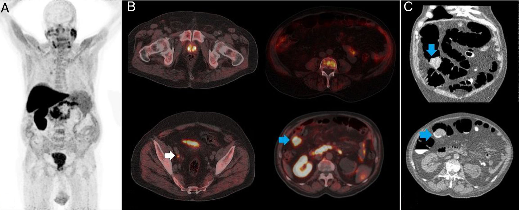
A 74-year-old man had a history of hormone-resistant, locally advanced prostate cancer. He was referred to our service due to suspicion of locoregional recurrence suggested in the control computed tomography (CT) scan that showed vesical trigone and left seminal vesicle involvement. Compromise was more doubtful at the level of 2 right pelvic regional lymph node chains. An 18F-choline PET/CT study was ordered for restaging (Fig. 1A and B) and identified a hypermetabolic prostate gland, along with metabolically positive right pararectal and external iliac adenopathies (white arrow), suggestive of malignancy, and also identified spinal bone infiltration. In addition, the imaging study showed hypermetabolic parietal thickening (30 mm) at the hepatic flexure (blue arrow), with a maximum standard uptake value (SUV) of 11.4, thus ruling out malignancy. Given the diagnosis of low-grade dysplastic villous adenoma made from the colonoscopic biopsy (Fig. 1C), right hemicolectomy was performed, revealing a well-differentiated, infiltrating adenocarcinoma (G1) that extended to the submucosa (pT1), under the villous adenoma. In the present case, 18F-choline PET/CT enabled the complete restaging of the prostatic lesion, as well as the location and diagnosis of a previously unsuspected neoplasm.
 Maximum intensity projection (MIP) 18F-choline PET/CT, (B) PET/CT axial views of the prostatic lesion, hypermetabolic adenopathies, and bone infiltration, as well as of the tumor of the colon, and (C) virtual colonoscopic biopsy, confirming colon tumor malignancy.")
No specific grants were received from public sector agencies, the business sector, or non-profit organizations in relation to this article.
Conflict of interestThe authors declare that there is no conflict of interest.
Ethical considerationsThe authors declare that they have followed the protocols of their work center on the publication of patient data, preserving patient anonymity at all times. Informed consent was not required for the publication of the present case because the article contains no personal data that could identify the patient.
Please cite this article as: Moreno-Ballesteros A, de la Riva-Pérez PA, Calvo-Morón MC. Detección de tumor metacrónico de colon mediante PET/TC con 18F-Colina en paciente con cáncer de próstata. Revista de Gastroenterología de México. 2021;86:435–436.

