
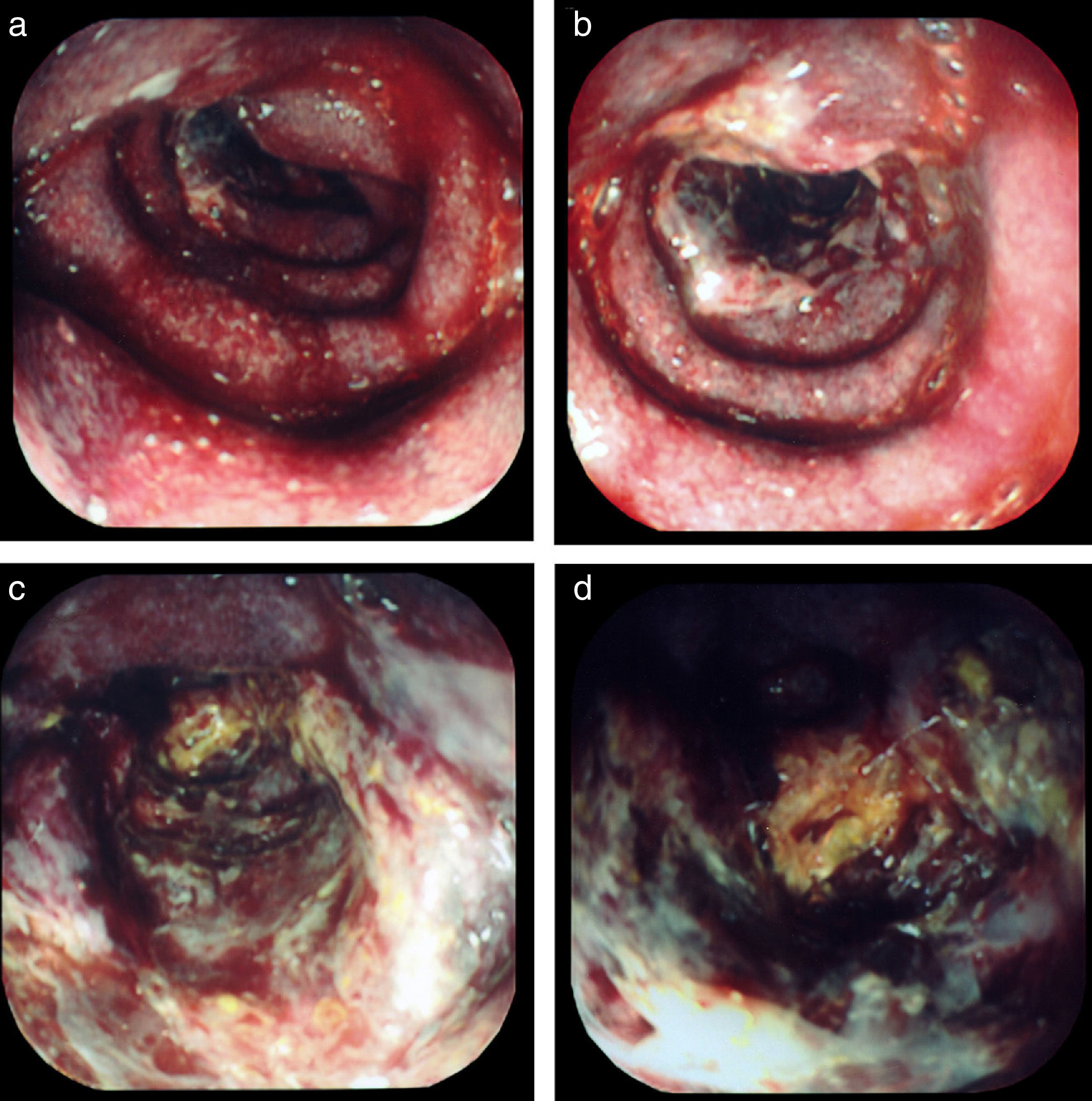
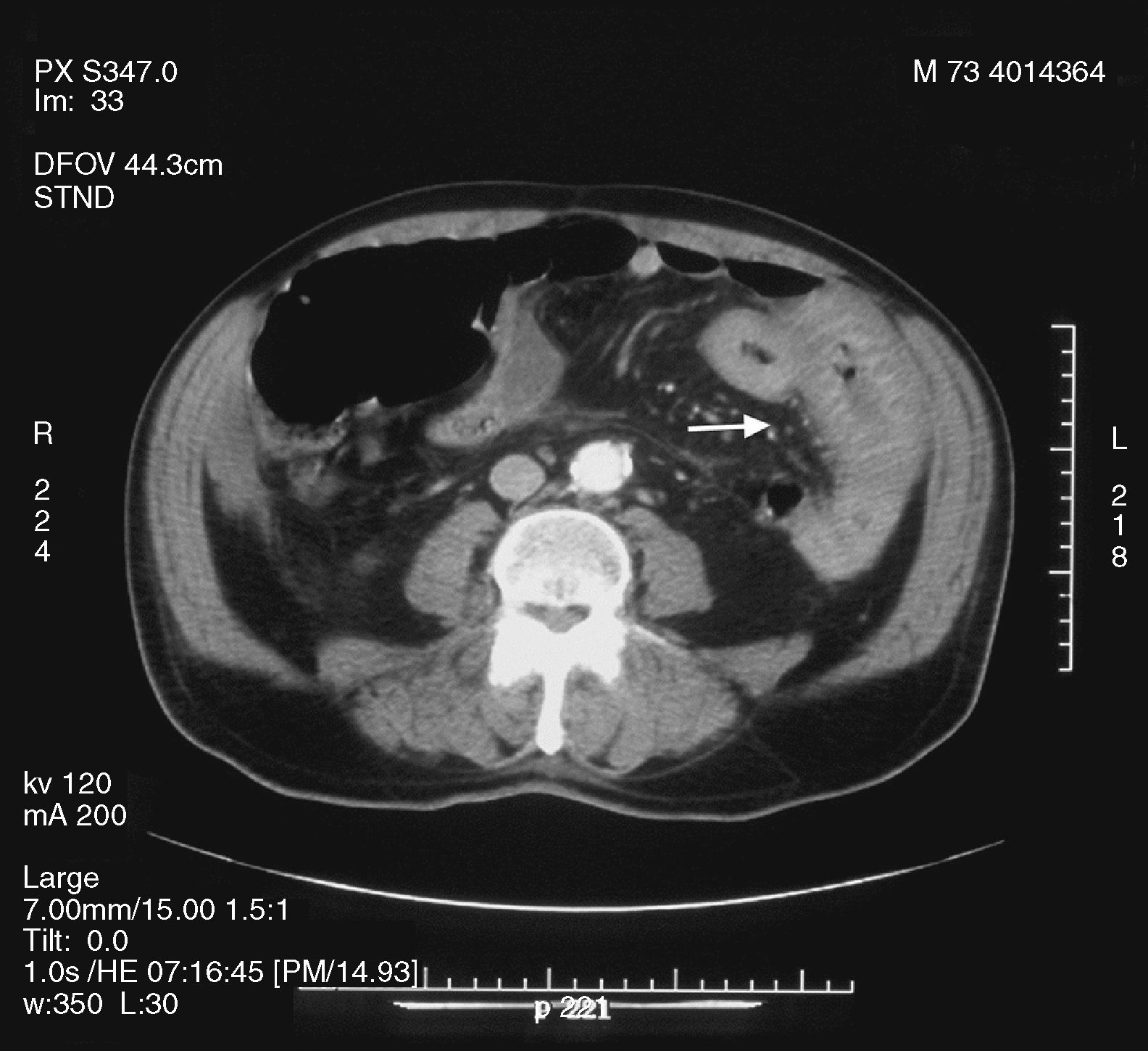
A 73-year-old man with hypertension and atrial fibrillation under warfarin was admitted with abdominal pain, distension, and dark blood hematemesis. He presented with anemia (Hb-8.4g/dL), prolonged prothrombin time and partial thromboplastin time with an INR >15. An endoscopy was performed. No lesions were identified, reporting only blood in the duodenum. A 150cm long pediatric colonoscope was introduced disclosing purple mucosa with ulcerations in the distal duodenum with spontaneous bleeding (Fig. 1a). As the endoscope passed into the jejunum, large and deeper ulcers were observed (Fig. 1b). Approximately 15cm distal to the Treitz angle (Fig. 1c), the bowel was extremely edematous presenting as circumferential ischemic necrosis. The ulcerations became deeper and about 25cm distal to the Treitz angle (Fig. 1d), there was complete luminal obstruction. The CT-scan showed a thickened wall of the distal duodenum and proximal jejunum, with edema and intramural hemorrhage (Fig. 2), without arterial or venous obstruction. Spontaneous small bowel hematoma is a rare complication of anticoagulant therapy. To the best of our knowledge, this is the first case of an intramural hematoma causing anticoagulant ileus, whose primary diagnosis was made through endoscopy.

.")
No financial support was received in relation to this article.
Conflict of interestThe authors declare that there is no conflict of interest.
Please cite this article as: Fonseca J, Meira T, Nunes A. Diagnóstico endoscópico de un hematoma intramural que se presentó como íleo por anticoagulantes. Revista de Gastroenterología de México. 2013;78:249–250.

