Journal Information
Vol. 88. Issue 4.
Pages 429-430 (October - December 2023)
Share
Share
Download PDF
More article options
Visits
802
Vol. 88. Issue 4.
Pages 429-430 (October - December 2023)
Clinical image in Gastroenterology
Full text access
Dysphagia lusoria
Disfagia lusoria
Visits
802
O. Gómez-Escudero
Corresponding author
octavio_gomezmd@yahoo.com.mx

Corresponding author at: Clínica de Gastroenterología, Endoscopía Digestiva y Motilidad Gastrointestinal “Endoneurogastro”, Hospital Ángeles Puebla, Puebla, Pue, Mexico.
Clínica de Gastroenterología, Endoscopía Digestiva y Motilidad Gastrointestinal “Endoneurogastro”, Hospital Ángeles, Puebla, Puebla, Mexico
This item has received
Article information
Full Text
Bibliography
Download PDF
Statistics
Figures (1)
Full Text

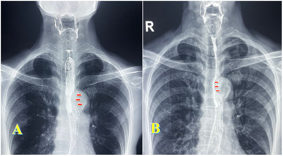
Dysphagia lusoria is a term utilized to describe dysphagia secondary to extrinsic vascular compression of the esophagus. The abnormal condition was described in 1794 by David Bayford in a 62-year-old female patient with dysphagia secondary to an aberrant right subclavian artery.1 It has also been associated with extrinsic compression of the aortic arch or one of its branches, with a prevalence of 0.5–1.8%.2,3 The word “lusoria” comes from the Latin phrase lusus naturae, which means “freak of nature”.3 The diagnostic method is a barium swallow test, and when there is doubt, angiotomography. Treatment depends on symptom severity and ranges from dietary modifications and alternating small bolus and liquid swallows to focusing on the cardiovascular disease causing the compression, which can include medical, surgical, or interventional radiology treatment, to obliterate or redirect the aberrant vessel.3,4 The case of an 81-year-old woman with intermittent retrosternal dysphagia to solids is presented herein. The videofluoroscopic swallow study shows extrinsic compression exactly at the level of the aortic arch (Fig. 1A and B), consistent with the diagnosis of dysphagia lusoria.

Figure 1.

(A) Videofluoroscopic swallow study showing extrinsic esophageal compression at the level of the aortic arch. (B) The same finding, with reduced caliber of barium passage at the level of the aortic arch.

(0.08MB).
Ethical considerations

The author declares that no experiments were conducted on animals or humans, that the present work maintains patient data anonymity. Informed consent was obtained from the patient for publishing this article. This document is in the possession of the corresponding author, meets the current bioethical research regulations.

Financial disclosure

No financial support was received in relation to this article.

Conflict of interest

The author declares that there is no conflict of interest.

References
[1]
N. Asherson.
David Bayford. His syndrome and sign of dysphagia lusoria.
Ann R Coll Surg Engl, 61 (1979), pp. 63-67
[2]
G.J. Carrizo, M.A. Marjani.
Dysphagia lusoria caused by an aberrant right subclavian artery.
Tex Heart Inst J, 31 (2004), pp. 168-171
[3]
B. Levitt, J.E. Richter.
Dysphagia lusoria: a comprehensive review.
Dis Esophagus, 20 (2007), pp. 455-460
[4]
A.L. Bennett, C. Cock, R. Heddle, et al.
Dysphagia lusoria: a late onset presentation.
World J Gastroenterol, 21 (2013), pp. 2433-2436

Please cite this article as: Gómez-Escudero O. Dysphagia lusoria. Rev Gastroenterol Mex. 2023;88:429–430.

Idiomas
Revista de Gastroenterología de México
Article options
Tools
es en
Política de cookies Cookies policy
Utilizamos cookies propias y de terceros para mejorar nuestros servicios y mostrarle publicidad relacionada con sus preferencias mediante el análisis de sus hábitos de navegación. Si continua navegando, consideramos que acepta su uso. Puede cambiar la configuración u obtener más información aquí. To improve our services and products, we use "cookies" (own or third parties authorized) to show advertising related to client preferences through the analyses of navigation customer behavior. Continuing navigation will be considered as acceptance of this use. You can change the settings or obtain more information by clicking here.