Journal Information
Vol. 79. Issue 4.
Pages 290-291 (October - December 2014)
Vol. 79. Issue 4.
Pages 290-291 (October - December 2014)
Clinical image in Gastroenterology
Open Access
Benign esophageal perforation resolved through the placement of a fully covered self-expandable metal stent
Perforación esofágica benigna resuelta mediante la colocación de una prótesis metálica autoexpandible totalmente cubierta
Visits
6640
E. Vázquez-Mora
Corresponding author
esfenoides12@hotmail.com

Corresponding author: Félix Cuevas 314 Depto. 701. Colonia del Valle, Mexico City CP 03100. Phone: +55 23 95 75 40.
, A. Farca-Belsaguy, D. Angulo-Molina, J.C. Salceda-Otero, D. Lozoya-González, F. Romero-Vallejo
Unidad de Endoscopia Avanzada, Centro Médico ABC, Mexico City, Mexico
This item has received

Under a Creative Commons license
Article information
Full Text
Download PDF
Statistics
Figures (4)
Show moreShow less
Full Text

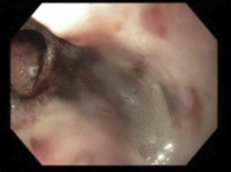
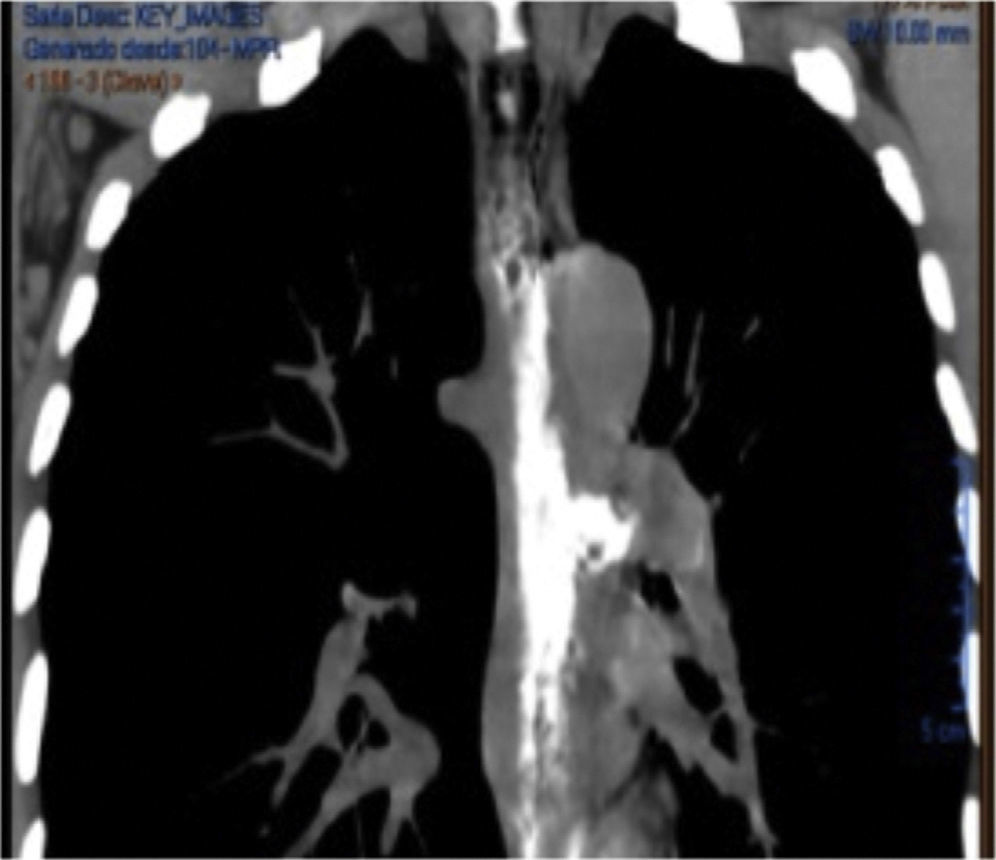
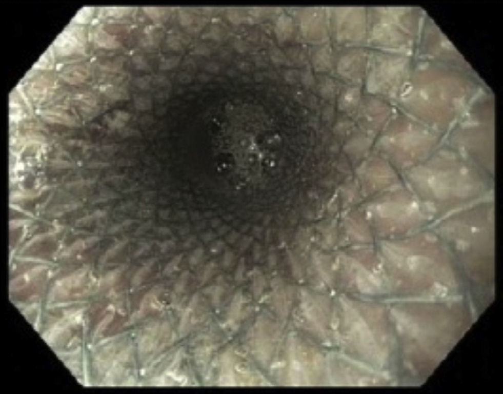
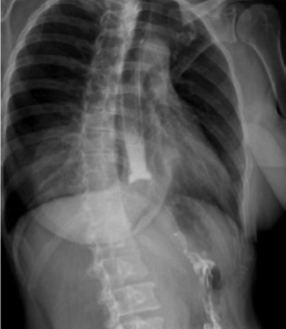
A 46-year-old woman presented with a history of 30-day nonsteroidal anti-inflammatory drug (NSAID) intake and dysphagia of 8-day progression. Her endoscopic study found a long (12cm) concentric stricture 22cm from the dentary arcade for which a gradual dilatation up to 15mm was performed with Savary-Gilliard dilators; the middle third of the esophagus was perforated during the second dilatation session and the lesion measured approximately 10mm in diameter (fig. 1). A chest computed tomography scan revealed pneumomediastinum and an image consistent with a perforation located at the middle third of the esophagus on the left lateral surface (fig. 2). Antibiotic therapy was begun and a 20 x 150mm fully covered self-expandable metal stent (Niti-S Esophageal Covered Stent, TaeWoong Medical,Seoul, Korea) was placed (fig. 3). A water-soluble contrast swallow (CS) was conducted and it showed adequate passage of the contrast medium through the stent with no evidence of leakage (fig. 4). Oral intake was restarted on the fifth day, the stent was removed 28 days later, and no solution of continuity was found in the esophagus.

Figure 1.

Esophageal perforation.

(0.06MB).
Figure 2.

The chest computed tomography scan with contrast medium showing pneumomediastinum and contrast material leakage at the left lateral surface of the esophagus.

(0.07MB).
Figure 3.

The fully covered esophageal metal stent occluding the entire perforation.

(0.09MB).
Figure 4.

The water-soluble contrast swallow (CS) study in which no leak into the mediastinum is observed.

(0.07MB).
Financial disclosure

No financial support was received in relation to this article.

Conflict of interest

The authors declare that there is no conflict of interest.

Please cite this article as: Vázquez-Mora E, Farca-Belsaguy A, Angulo-Molina D, Salceda-Otero JC, Lozoya-González D, Romero-Vallejo F. Benign esophageal perforation resolved through the placement of a fully covered self-expandable metal stent. Revista de Gastroenterología de México. 2014;79:290–291.

Copyright © 2014. Asociación Mexicana de Gastroenterología
Idiomas
Revista de Gastroenterología de México
Article options
Tools
es en
Política de cookies Cookies policy
Utilizamos cookies propias y de terceros para mejorar nuestros servicios y mostrarle publicidad relacionada con sus preferencias mediante el análisis de sus hábitos de navegación. Si continua navegando, consideramos que acepta su uso. Puede cambiar la configuración u obtener más información aquí. To improve our services and products, we use "cookies" (own or third parties authorized) to show advertising related to client preferences through the analyses of navigation customer behavior. Continuing navigation will be considered as acceptance of this use. You can change the settings or obtain more information by clicking here.