Journal Information
Vol. 86. Issue 3.
Pages 309-310 (July - September 2021)
Visits
4474
Vol. 86. Issue 3.
Pages 309-310 (July - September 2021)
Clinical image in Gastroenterology
Open Access
Acute pancreatitis manifesting in the skin: Pancreatic panniculitis
La pancreatitis aguda se manifiesta en la piel: paniculitis pancreática
Visits
4474
M.M. Díaz-Alcázara,
Corresponding author
mmardiazalcazar@gmail.com

Corresponding author. Avenida de la Investigación, s/n,18016 Granada, Spain. Tel.: +0034 958 02 30 00.
, J.L. López-Hidalgob, J. Aneiros-Fernándezb
a Unidad de Gestión Clínica de Aparato Digestivo, Hospital Universitario Clínico San Cecilio, Granada, Spain
b Unidad Provincial de Anatomía Patológica de Granada, Hospital Universitario Clínico San Cecilio, Granada, Spain
This item has received

Under a Creative Commons license
Article information
Full Text
Download PDF
Statistics
Figures (2)
Full Text

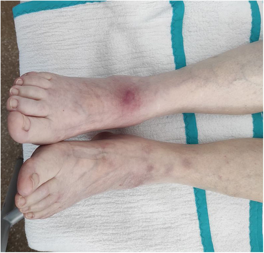
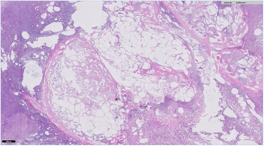
An 83-year-old woman was admitted for acute pancreatitis after consultation for epigastralgia and hyperamylasemia (13,370 U/l; normal range 20-120). Two days later, she presented with nonulcerated, painful, erythematous-purplish subcutaneous nodules on her legs and arms (Fig. 1). Ultrasound revealed inflammation of the subcutaneous cell tissue. A nodule was biopsied and confirmed pancreatic panniculitis (Fig. 2). Pancreatic panniculitis, or pancreatic fat necrosis, is a rare skin manifestation (< 3%) of pancreatic diseases. It can precede the diagnosis of pancreatic pathology, and so pancreatic disease screening should be carried out in patients that present with no abdominal symptomatology. Pancreatic panniculitis is clinically indistinguishable from other types of panniculitis, but the histologic findings are pathognomonic: lobular panniculitis with extensive necrosis of adipocytes and “ghost cells” (adipocytes that have no nucleus and a fine, granular, basophilic deposit due to calcium accumulation). Progression parallels that of pancreatic disease.

Figure 1.

Nonulcerated, erythematous-purplish subcutaneous nodules on the lower limbs.

(0.11MB).
Figure 2.

Histologic slice with hematoxylin-eosin staining at x50 magnification, showing lobular panniculitis with extensive necrosis of adipocytes and adipocytes that have no nucleus and a fine, granular, basophilic deposit.

(0.13MB).
Ethical considerations

The authors declare that:

  • ˗

    Informed consent was requested of the patient to receive treatment or participate in the study described.

  • ˗

    The Clinical Image meets the current bioethics research regulations and was authorized by the ethics committee of theHospital Universitario Clínico San Cecilio.

  • ˗

    This article contains no personal information that could identify the patient.

  • ˗

    No experiments were conducted on animals or humans.

Financial disclosure

No financial support was received in relation to this article.

Conflict of interest

The authors declare that there is no conflict of interest.

Please cite this article as: Díaz-Alcázar MM, López-Hidalgo JL, Aneiros-Fernández J. La pancreatitis aguda se manifiesta en la piel: paniculitis pancreática. Rev Gastroenterol Méx. 2021;86:309–310.

Copyright © 2021. Asociación Mexicana de Gastroenterología
Idiomas
Revista de Gastroenterología de México
Article options
Tools
es en
Política de cookies Cookies policy
Utilizamos cookies propias y de terceros para mejorar nuestros servicios y mostrarle publicidad relacionada con sus preferencias mediante el análisis de sus hábitos de navegación. Si continua navegando, consideramos que acepta su uso. Puede cambiar la configuración u obtener más información aquí. To improve our services and products, we use "cookies" (own or third parties authorized) to show advertising related to client preferences through the analyses of navigation customer behavior. Continuing navigation will be considered as acceptance of this use. You can change the settings or obtain more information by clicking here.