Journal Information
Vol. 79. Issue 2.
Pages 145-146 (April - June 2014)
Share
Share
Download PDF
More article options
Vol. 79. Issue 2.
Pages 145-146 (April - June 2014)
Clinical image in Gastroenterology
Open Access
A rare cause of intussusception in the adult: Intestinal bezoar
Excepcional causa de intususcepción en el adulto: bezoar intestinal
Visits
8585
P. Caleroa,
Corresponding author
pcalerogarcia@medicos.com

Corresponding author: Servicio de Cirugía General y Digestiva del Hospital de Hellín. Hospital de Hellín. Calle Juan Ramón Jiménez, 40. CP 02400 Hellín, Albacete. Tel.: +34658432088; fax: +34967597202.
, M. Scortechinia, J. Valienteb
a General and Gastrointestinal Surgery Service, Hospital de Hellín, Hellín, Albacete, Spain
b General and Gastrointestinal Surgery Service Management, Hospital de Hellín, Hellín, Albacete, Spain
This item has received

Under a Creative Commons license
Article information
Full Text
Download PDF
Statistics
Figures (4)
Show moreShow less
Full Text

Intestinal intussusception is a rare cause of bowel obstruction in the adult, and no such one produced by a bezoar has been described in the medical literature.

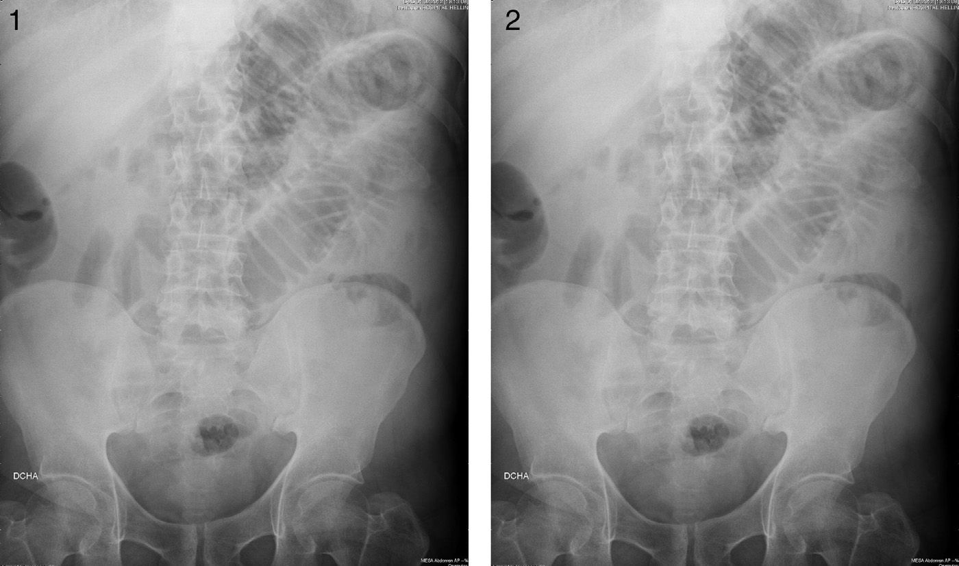
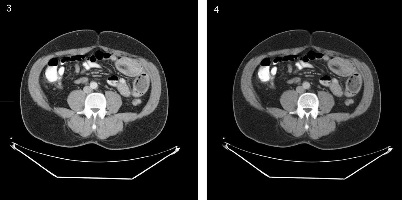
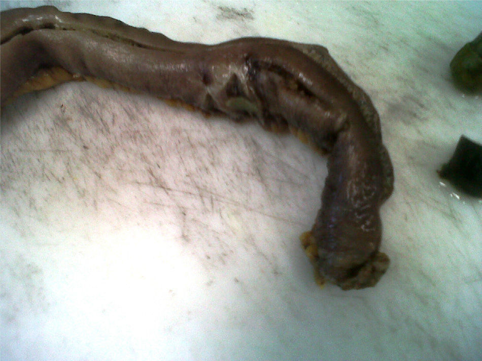
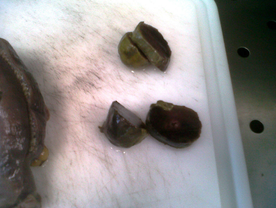
A 49-year-old man with an unremarkable past medical history came to the Emergency Department complaining of abdominal pain in the epigastrium of 3-4 day progression, associated with a reduced number and quantity of daily bowel movements. Physical examination was consistent with bowel obstruction. A plain abdominal film revealed segment dilation up to the jejunum (Figs. 1 and 2). An abdominal computed axial tomography (CAT) scan identified an oval-shaped lesion that appeared to be an intestinal invagination in the right flank (Figs. 3 and 4). Given the diagnosis of bowel obstruction, emergency surgery was performed. Mechanical ileus of the small bowel in the mid jejunum due to invagination was observed, along with an intestinal bezoar that measured 6 x 4 x 3cm with 2 invaginated zones and ulcerated serous membrane. About 40cm of the small bowel was resected and an end-to-end anastomosis was performed (Figs. 5 and 6). Postoperative progression was satisfactory.

Figures 1 and 2.

Plain x-ray images showing small bowel obstruction data.

(0.13MB).
Figures 3 and 4.

Computed axial tomography images in which the segment involved in the intussusception can be seen.

(0.11MB).
Figure 5.

Image of the surgical specimen showing the ischemia produced by the bezoar.

(0.13MB).
Figure 6.

The sectioned bezoar.

(0.13MB).
Financial disclosure

No financial support was received in relation to this article.

Conflict of interest

The authors declare that there is no conflict of interest.

Please cite this article as: Calero P, Scortechini M, Valiente J. Excepcional causa de intususcepción en el adulto: bezoar intestinal. Revista de Gastroenterología de México. 2014;79:145–146.

Institute where study was carried out: Hospital de Hellín, Albacete, Spain.

Copyright © 2014. Asociación Mexicana de Gastroenterología
Idiomas
Revista de Gastroenterología de México
Article options
Tools
es en
Política de cookies Cookies policy
Utilizamos cookies propias y de terceros para mejorar nuestros servicios y mostrarle publicidad relacionada con sus preferencias mediante el análisis de sus hábitos de navegación. Si continua navegando, consideramos que acepta su uso. Puede cambiar la configuración u obtener más información aquí. To improve our services and products, we use "cookies" (own or third parties authorized) to show advertising related to client preferences through the analyses of navigation customer behavior. Continuing navigation will be considered as acceptance of this use. You can change the settings or obtain more information by clicking here.