

Case
A 66-year-old man with an unremarkable past medical history was admitted to the hospital with symptoms of abdominal pain of 36-hour progression suggestive of acute appendicitis. Laboratory work-up reported: 21,500 leukocytes/cc, neutrophils 84.8%, and the remaining tests were normal. A plain anteroposterior abdominal x-ray showed the presence of 2 radio-opaque, filiform, sharp-ended, metallic objects measuring approximately 5 cm and located in the lower right abdominal quadrant (Fig. 1). During the medical interview, the patient denied having swallowed any type of foreign body in the previous days or months. The diagnosis of intestinal perforation secondary to foreign bodies versus appendicitis secondary to foreign bodies was established. Exploratory laparotomy revealed an approximately 9 cm retrocecal appendix perforated at its middle third section by the protruding tip of a straight pin, with its body and a second pin inside the appendicular lumen. Appendectomy was performed with no complications. The presence of the two foreign bodies inside the appendicular lumen was corroborated in the operating room through x-ray of the specimen (Fig. 2). The histologic report of the specimen was acute appendicitis with abscess and the presence of 2 straight pins in the lumen (Fig. 3). The patient progressed satisfactorily and was released 48 hours after surgery.
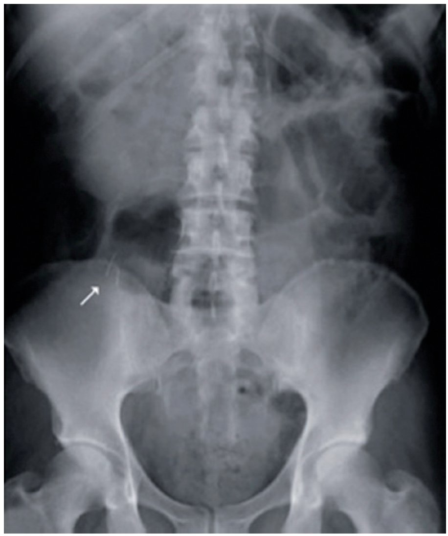
Figure 1. AP x-ray of the 66-year-old male patient showing the pins in the appendicular lumen.

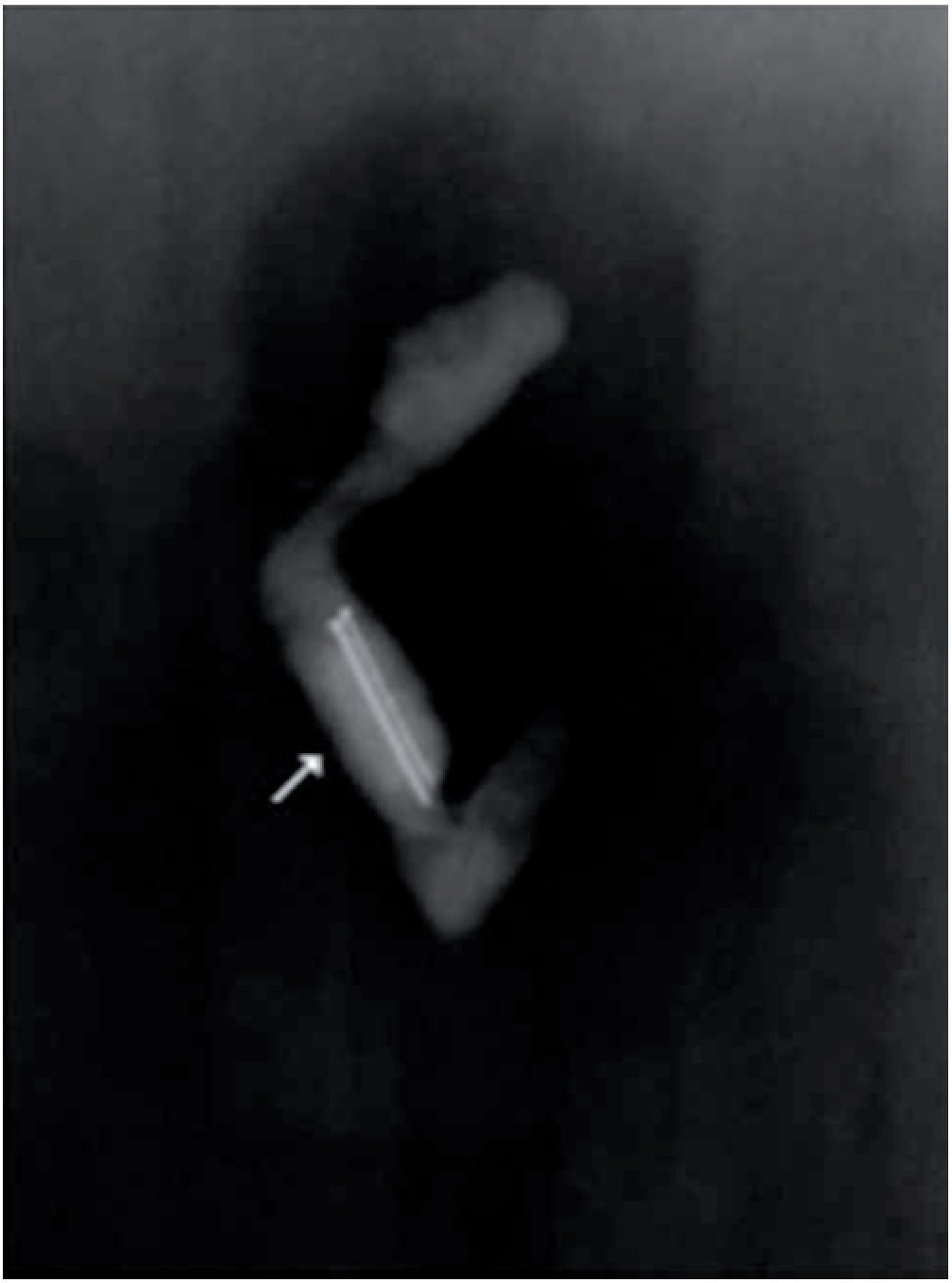
Figure 2. X-ray of the surgical specimen showing the 2 pins inside the appendicular lumen.
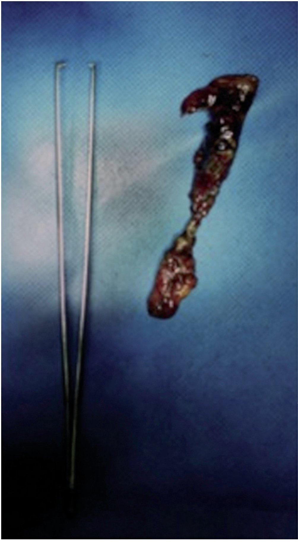
Figure 3. Cecal appendix with fibrinous and purulent peritonitis and necrosis. The pins in its interior are not shown.
qPlease cite this article as: Valenzuela¿Salazar C, et al. Apendicitis causada por alfileres. Revista de Gastroenterología de México. 2013;78:45-6.
* Corresponding author at:
Hospital General Dr. Manuel Gea González, Departamento de Cirugía General y Endoscópica, Calzada de Tlalpan, 4800 6.o piso, Col Sección XVI, Delegación Tlalpan, CP 14080, México D.F., Mexico.
Tel: +(52) 55 40003000 ext 3329; Fax: +(52) 55 40003000 ext 3329; Cell phone: +(52) 55 19484679.
Email address: carlosvalenzuelas@gmail.com (C. Valenzuela-Salazar).

