
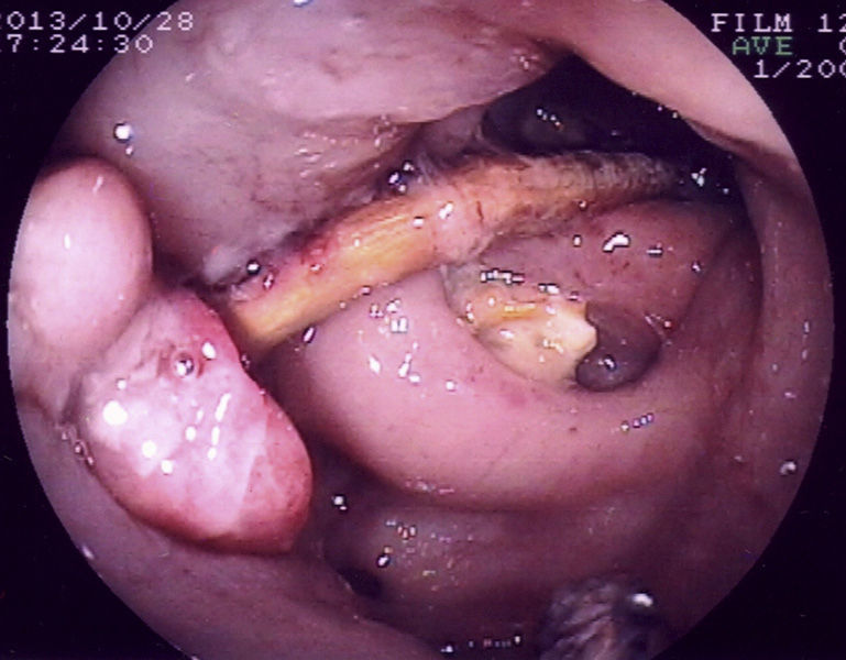
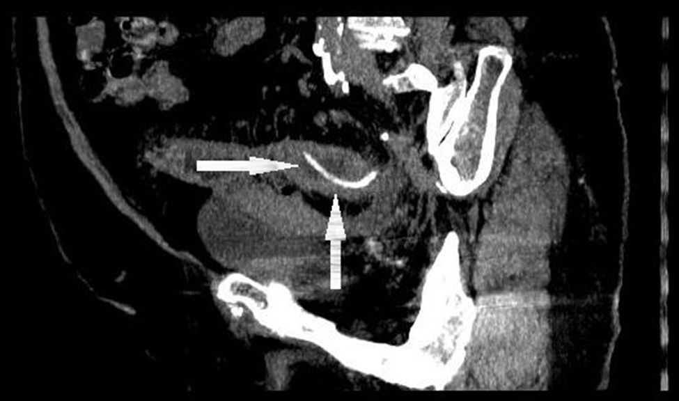
An asymptomatic 79-year-old woman, with an unremarkable past medical history, came to the endoscopy service for a programmed colonoscopy, as a screening procedure due to a family history of colorectal cancer. The endoscopy (fig. 1) identified a large quantity of wide-mouthed diverticular orifices at the level of the sigmoid colon and a smooth, curved foreign body (chicken bone) penetrating the mucosa, surrounded by an important inflammatory reaction. An unsuccessful attempt was made to extract it through traction. An abdominal computed tomography scan (fig. 2) revealed an elongated, L-shaped bone, penetrating the wall of the sigmoid colon, producing a thickening of the right pelvic fascia (inflammatory/fibrous reaction). The patient refused surgical treatment and asked to be released. Two months later, the patient returned for consultation after spontaneously expulsing the bone through bowel movements. There are many published articles in relation to perforation of sigmoid colon diverticula by foreign objects, the most common of which are fish bones. However, asymptomatic presentation followed by spontaneous expulsion is an anecdotal case.
 impacted in the diverticular orifice of the sigmoid colon, with an intense inflammatory reaction.")
 penetrating the wall of the sigmoid colon, with pelvic fascia thickening.")
The authors declare that no experiments on humans or animals were carried out in relation to this study.
Data confidentialityThe authors declare that no patient data appear in this article.
Right to privacy and informed consentThe authors declare that no patient data appear in this article.
Financial disclosureNo financial support was received in relation to this study/article.
Conflict of interestThe authors declare that there is no conflict of interest.
Please cite this article as: Domínguez-Jiménez JL, Jaén-Reyes MT. Perforación de divertículo de colon sigmoide por hueso de pollo. Revista de Gastroenterología de México. 2015;80:107–108.

