Journal Information
Vol. 83. Issue 3.
Pages 346-347 (July - September 2018)
Vol. 83. Issue 3.
Pages 346-347 (July - September 2018)
Clinical image in Gastroenterology
Open Access
Gastric obstruction secondary to an intragastric balloon
Obstrucción gástrica secundaria a balón intragástrico
Visits
6346
D. Rubio Solísa,
Corresponding author
diegors.radiologia@gmail.com

Corresponding author. Radiodiagnóstico, Hospital Universitario Central de Asturias, Av. de Roma, s/n, 33011 Oviedo, Asturias, Spain. Phone: 628489470.
, S. Sánchez Garcíab
a Servicio de Radiodiagnóstico, Hospital Universitario Central de Asturias, Oviedo, Asturias, España
b Servicio de Radiodiagnóstico, Hospital Universitario de San Agustín, Avilés, Asturias, España
This item has received

Under a Creative Commons license
Article information
Full Text
Download PDF
Statistics
Figures (3)
Show moreShow less
Full Text

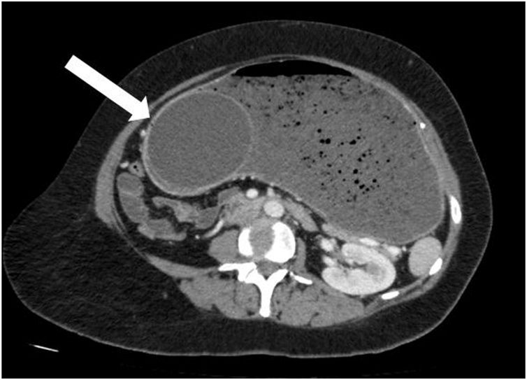
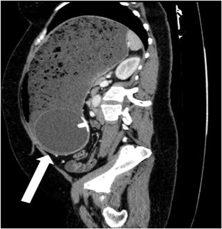
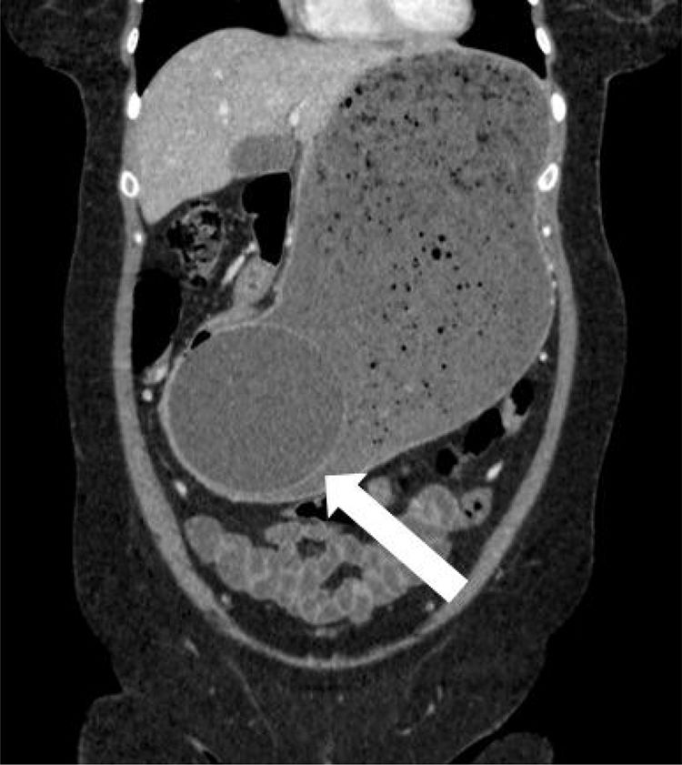
A 45-year-old woman came to the emergency service due to 3 days of ongoing vomiting and significant generalized and progressive abdominal distension. Her past medical history was unremarkable, except for the placing of an intragastric balloon 3 weeks prior. Plain abdominal x-ray revealed an occupied abdomen and bowel loop displacement toward the periphery. A computed axial tomography scan identified severe dilation of the gastric corpus, along with the intragastric balloon (white arrow), which was embedded in the antral region, causing the gastric outlet obstruction (Figure 1 in the axial plane, Figure 2 in the sagittal plane, and Figure 3 in the coronal plane). According to the medical literature, frequency of overall complications from intragastric balloon placement reaches 2.6% and obstruction is one of the most common causes in the early postoperative period, with a frequency of 0.8%. First, a nasogastric tube was placed in our patient to relieve the distension, after which the obstruction was resolved through endoscopic removal of the balloon. After the intervention, the suggested cause was the progression of the balloon and its lodging in the antrum of the stomach, with no excessive filling of that gastric region.

Figure 1.

Oblique axial CT image showing the intragastric balloon embedded in the antrum of the stomach.

(0.05MB).
Figure 2.

Oblique sagittal CT image showing the intragastric balloon embedded in the antrum of the stomach and the great dilation of the entire stomach.

(0.07MB).
Figure 3.

Oblique coronal CT image showing the same findings.

(0.06MB).
Financial disclosure

No financial support was received in relation to this study/article.

Conflict of interest

The authors declare that there is no conflict of interest.

Please cite this article as: Rubio Solís D, Sánchez García S. Obstrucción gástrica secundaria a balón intragástrico. Revista de Gastroenterología de México. 2018;83:346–347.

Idiomas
Revista de Gastroenterología de México
Article options
Tools
es en
Política de cookies Cookies policy
Utilizamos cookies propias y de terceros para mejorar nuestros servicios y mostrarle publicidad relacionada con sus preferencias mediante el análisis de sus hábitos de navegación. Si continua navegando, consideramos que acepta su uso. Puede cambiar la configuración u obtener más información aquí. To improve our services and products, we use "cookies" (own or third parties authorized) to show advertising related to client preferences through the analyses of navigation customer behavior. Continuing navigation will be considered as acceptance of this use. You can change the settings or obtain more information by clicking here.