Journal Information
Vol. 84. Issue 3.
Pages 398-399 (July - September 2019)
Vol. 84. Issue 3.
Pages 398-399 (July - September 2019)
Clinical image in Gastroenterology
Open Access
Fatal pancreatic inflammation: Extensive emphysematous pancreatitis diagnosed by computed tomography
Inflamación pancreática letal: pancreatitis enfisematosa extensa diagnosticada por tomografía computarizada
Visits
5637
S. Junquera-Olay
Corresponding author
junqueraolays@gmail.com

Corresponding author. Servicio de Radiodiagnóstico, Complejo Hospitalario Universitario de Santiago de Compostela, Avenida Da Choupana s/n, 15706 Santiago de Compostela, A Coruña, Spain.
, S. Baleato-González, R. García-Figueiras
Servicio de Radiodiagnóstico, Complejo Hospitalario Universitario de Santiago de Compostela, Santiago de Compostela, A Coruña, Spain
This item has received

Under a Creative Commons license
Article information
Full Text
Download PDF
Statistics
Figures (4)
Show moreShow less
Full Text

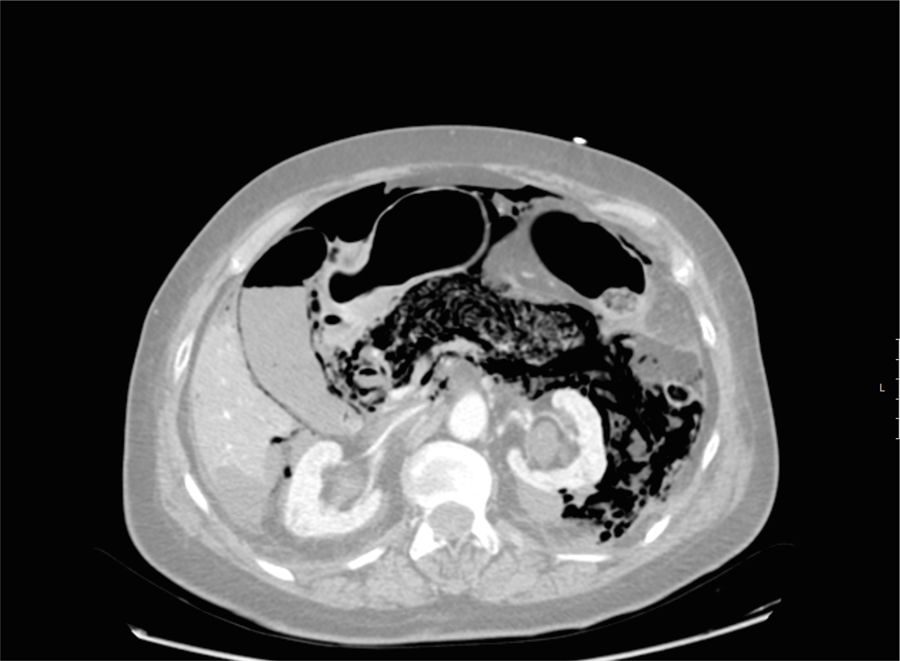
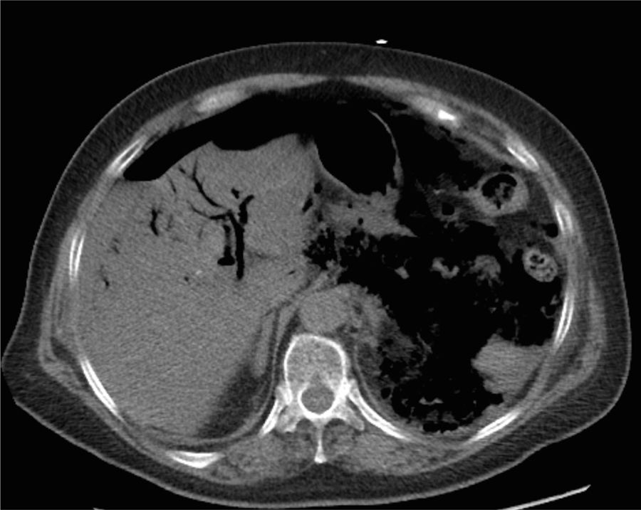
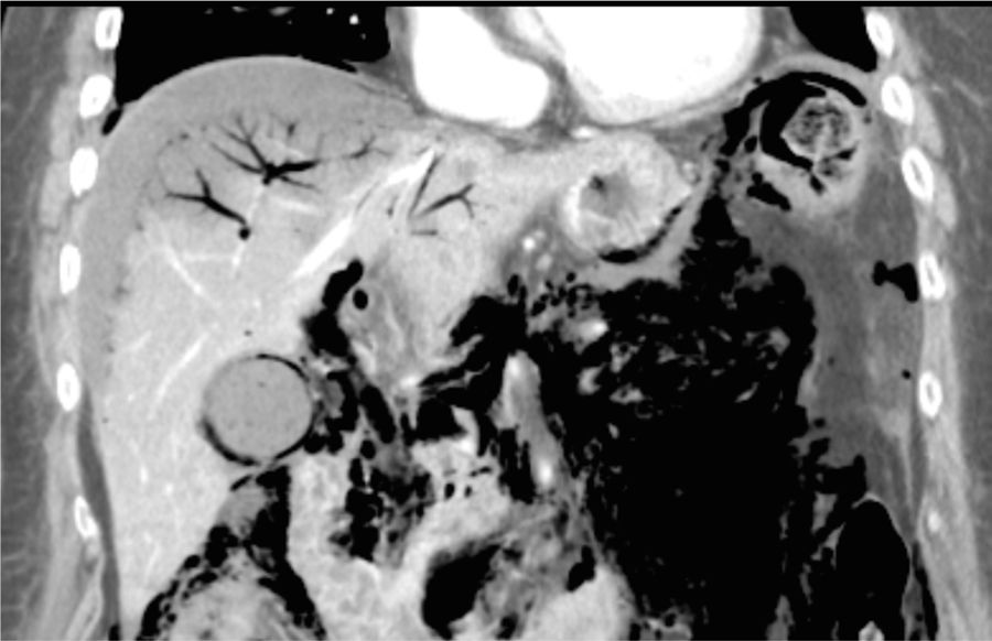
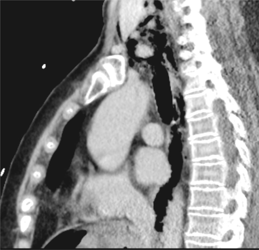
An 82-year-old diabetic woman presented with clinical symptoms of vomiting and abdominal pain. At the emergency service, blood test results showed serum amylase and lipase levels of 2,141 IU/l and 12,864 IU/l, respectively. The patient had no history of a previous biliary endoscopic procedure. After a few hours, she began to present with signs of shock. An abdominal-pelvic CT scan was carried out to rule out complications. In the axial view at the level of the pancreas and the upper poles of the kidneys, emphysema in the pancreatic parenchyma was identified that extended into the left perirenal and mesenteric fat, associated with ascitic fluid (fig. 1). Pneumobilia was identified at the hepatic level, along with the presence of a large quantity of intra-abdominal gas (fig. 2). In the coronal reconstruction, gas in the gallbladder at both the parietal and endoluminal levels was better visualized, as well as in the biliary tract and the portal branches (fig. 3). The process was so extensive that it caused pneumoperitoneum and it spread into the periesophageal mediastinum, resulting in emphysema of the subcutaneous tissue of the neck (fig. 4).

Figure 1.

Axial CT view at the level of the pancreas, gallbladder, and upper poles of the kidneys showing emphysema in the pancreatic parenchyma that extends into the left perirenal and mesenteric fat, associated with ascitic fluid and pneumoperitoneum.

(0.05MB).
Figure 2.

Axial view at the hepatic level, showing pneumobilia and the presence of a large quantity of intra-abdominal gas.

(0.07MB).
Figure 3.

Coronal section of the same patient, with better visualization of the gas in the gallbladder, at both the parietal and endoluminal levels, and in the biliary tract and the portal branches.

(0.06MB).
Figure 4.

The process was so extensive that it caused pneumoperitoneum and spread into the periesophageal mediastinum, causing emphysema of the subcutaneous tissue of the neck.

(0.07MB).

We were facing a case of emphysematous pancreatitis, a rare and severe complication of acute pancreatitis, with a mortality rate above 35%. It is characterized by the presence of gas in and/or around the pancreatic gland due to a necrotizing infection caused by gas-producing bacteria. The patient was admitted to the intensive care unit, where she presented with multiple organ failure, and died 6hours after having the imaging study and 34hours after having arrived at the emergency service. The speed with which the disease was established could be explained by the patient's history of diabetes mellitus, which could have attenuated the symptomatology, delaying the diagnosis, or by the possibility that the pancreatitis was accompanied by perforation of a hollow viscus, albeit no definitive radiologic signs confirming that hypothesis were identified.

Financial disclosure

No financial support was received in relation to this article.

Conflict of interest

The authors declare that there is no conflict of interest.

Please cite this article as: Junquera-Olay S, Baleato-González S, García-Figueiras R. Inflamación pancreática letal: pancreatitis enfisematosa extensa diagnosticada por tomografía computarizada. Revista de Gastroenterologí a de México. 2019;84:398–399.

Copyright © 2018. Asociación Mexicana de Gastroenterología
Idiomas
Revista de Gastroenterología de México
Article options
Tools
es en
Política de cookies Cookies policy
Utilizamos cookies propias y de terceros para mejorar nuestros servicios y mostrarle publicidad relacionada con sus preferencias mediante el análisis de sus hábitos de navegación. Si continua navegando, consideramos que acepta su uso. Puede cambiar la configuración u obtener más información aquí. To improve our services and products, we use "cookies" (own or third parties authorized) to show advertising related to client preferences through the analyses of navigation customer behavior. Continuing navigation will be considered as acceptance of this use. You can change the settings or obtain more information by clicking here.