
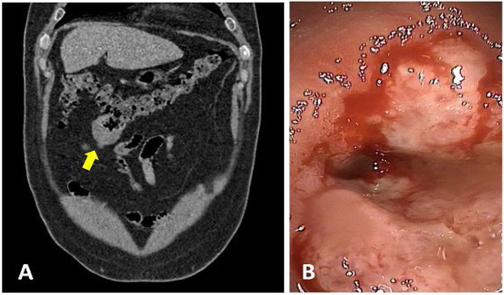
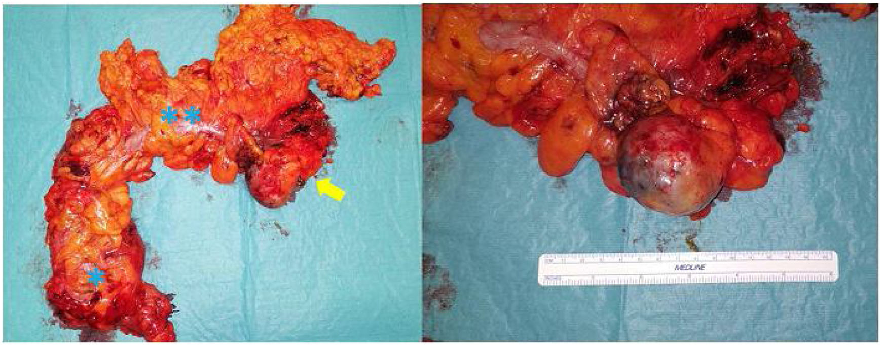
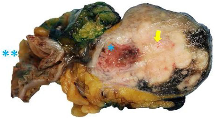
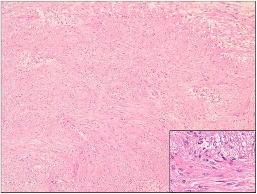
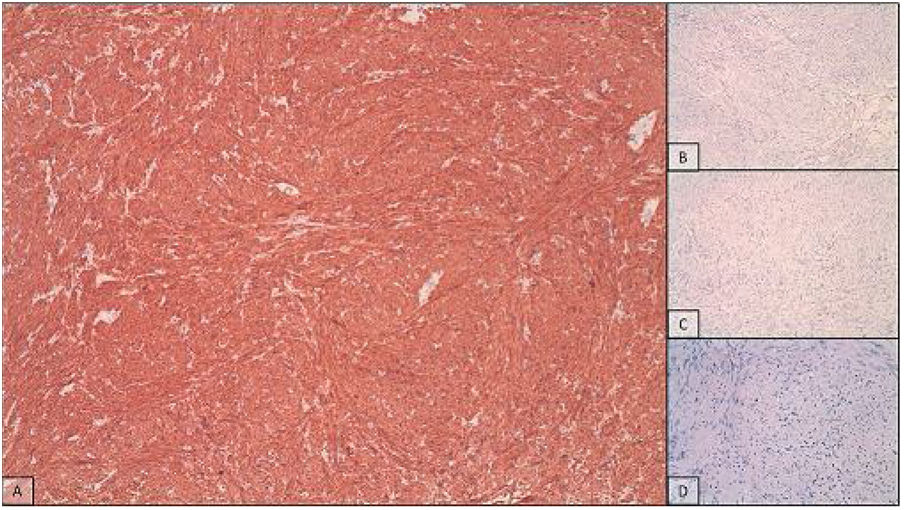
Colonic leiomyomas of the colon are infrequent entities, the most common of which are sessile and intraluminal lesions that can be endoscopically resected. They arise from the muscularis propria or the muscularis mucosae, but can invade the serosa, intestinal lumen, or both. A 60-year-old man had the incidental finding, on a computerized tomography (CT) scan, of a 5 cm lesion, with a diverticular morphology, in the transverse colon (Fig. 1A). Colonoscopy identified a strictured and ulcerated lesion that impeded the passage of the colonoscope, but histologically, was benign (Fig. 1B). Surgical treatment revealed a dumbbell-shaped lesion, dependent on the antimesenteric border of the colon. Laparoscopic extended right hemicolectomy was performed (Fig. 2). Pathologic anatomy described a dumbbell lesion with intraluminal and extraluminal growth (Fig. 3) and cellular proliferation arranged in fascicles (Fig. 4). Immunohistochemistry revealed cells with intense and diffuse smooth muscle actin expression (Fig. 5). The majority of these tumors are asymptomatic, such as in our patient, but they can produce pain, bleeding, or obstruction. They are benign tumors, but can recur and metastasize, making their follow-up necessary.
 CT coronal view, showing the dumbbell-shaped lesion (arrow) of the transverse colon. B) Colonoscopic image, showing a friable and strictured lesion.")
, transverse colon (**), and dumbbell-shaped leiomyoma (arrow) can be seen.")
 and intraluminal growth, partially stricturing the intestinal lumen (*), resulting in its characteristic shape. The specimen also shows the normal section of the intestinal lumen (**).")

 and are negative for C-kit (B) and S100 (C), with a low Ki67 proliferation index (D).")
The authors declare that no experiments were conducted on humans or animals for the present article, that they have followed the protocols of their work center on the publication of patient data, and that they have preserved patient confidentiality and anonymity at all times. Informed consent was requested from the patient for the surgical intervention and it included a section stating the possibility of utilizing images or clinical data for scientific purposes.
Financial disclosureNo financial support was received in relation to this article.
Conflict of interestThe authors declare that there is no conflict of interest.
Please cite this article as: Ruiz de la Hermosa A, Roldán-Cortés D, Paseiro-Crespo G. Leiomioma «dumbbell» de colon transverso. Rev Gastroenterol Méx. 2022;87:106–107.

