Journal Information
Vol. 82. Issue 2.
Pages 179-180 (April - June 2017)
Vol. 82. Issue 2.
Pages 179-180 (April - June 2017)
Clinical image in Gastroenterology
Open Access
“Downhill” esophageal varices secondary to superior vena cava thrombosis and abnormal factor V
Varices esofágicas en Downhill secundarias a trombosis de vena cava superior por déficit de factor V
Visits
8246
A.J. Gómez-Aldanaa,
Corresponding author
andresgomezmd@hotmail.com

Corresponding author. Departamento de Medicina Interna, Hospital Universitario San Ignacio, Cra. 7N.° 40-62, Bogotá, Colombia. Tel.: +7 1 5946161.
, M.A. Gómez-Zuletab
a Fundación Santa Fe de Bogotá, Servicio de Endoscopia, Hospital Universitario San Ignacio, Bogotá, Colombia
b Hospital El Tunal, Servicio de Gastroenterología, Universidad Nacional de Colombia, Bogotá, Colombia
This item has received

Under a Creative Commons license
Article information
Full Text
Bibliography
Download PDF
Statistics
Figures (3)
Show moreShow less
Full Text

Since the first description of “Downhill” varices,1 or varices in the proximal esophagus, by Felson and Lessure in 1964, there have been only about 80 articles on the theme in the literature. The appearance of this phenomenon in the proximal third of the esophagus arises from obstruction of the blood flow from the superior vena cava, proximal to the azygos vein, forcing the veins to drain through the mediastinal collateral vessels that appear in the proximal esophagus.2–4 Lung cancer, lymphoma, and mediastinal metastases are among the malignant causes of this entity5 and goiter, congenital or valvular heart disease, thymoma, and systemic vasculitides, such as Behcet's disease, are included among its benign causes. These varices can also be secondary to procedures such as pacemaker insertion or hemodialysis access.3,4 Hypercoagulability disorders are another etiology, with one case reported in the literature.2 We describe herein a case of “Downhill” esophageal varices secondary to a hypercoagulability disorder.

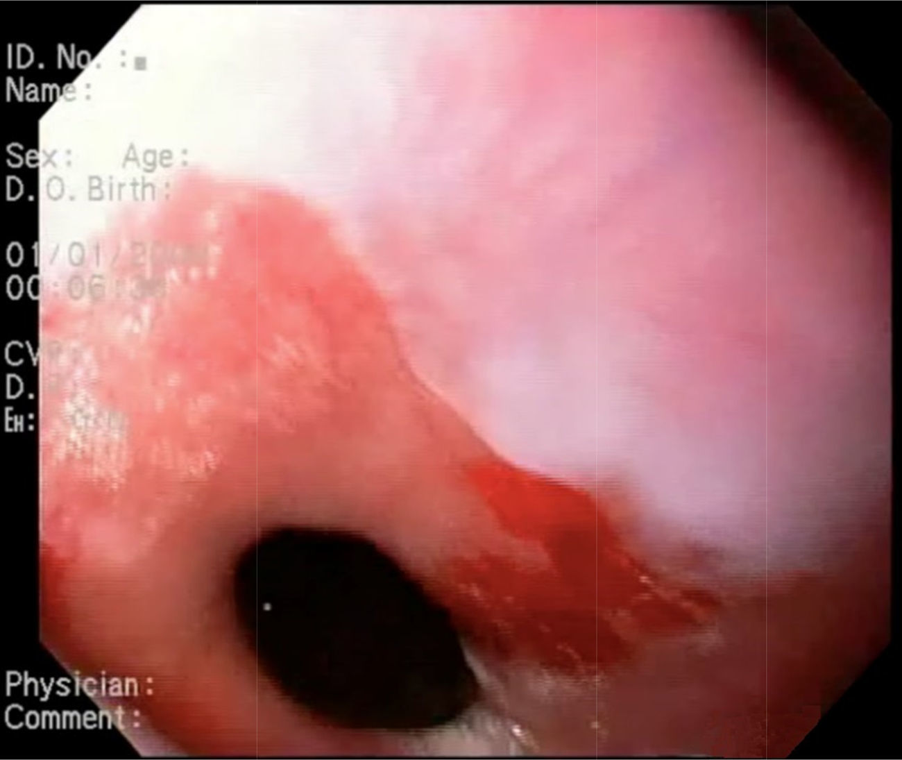
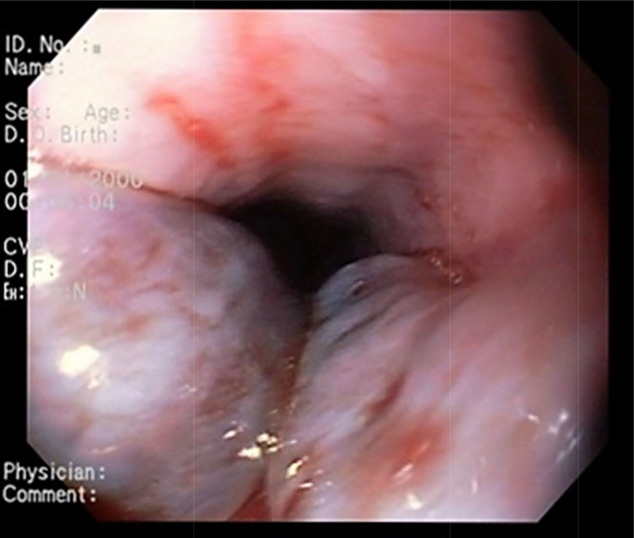
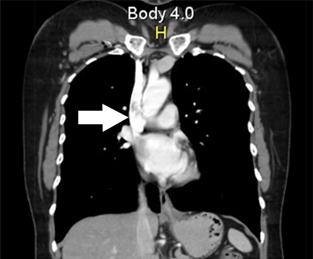
A 53-year-old woman with no remarkable past history was admitted to the emergency department due to hematemesis with no abdominal pain of 2-day progression. Physical examination revealed a heart rate of 90, blood pressure of 100/60, and rectal examination was positive for melena. At endoscopy, an unaltered gastroesophageal junction was observed (fig. 1), as well as 2 venous dilations in the proximal third of the esophagus with no evidence of active bleeding (fig. 2), suggestive of Downhill varices. A computed tomography chest scan showed an image of a thrombus in the superior vena cava (fig. 3). Anticoagulation management was begun with enoxaparin. Control endoscopy was carried out at 72h that revealed a reduction in the size of the esophageal varices. Hypercoagulability studies were begun, with normal results, except for the factor V Leiden mutation.

Figure 1.

Unaltered gastroesophageal junction.

(0.11MB).
Figure 2.

Venous dilations in the proximal third of the esophagus, with no evidence of active bleeding.

(0.12MB).
Figure 3.

Chest CT showing the thrombus in the superior vena cava.

(0.11MB).
Ethical disclosuresProtection of human and animal subjects

The authors declare that the procedures followed were in accordance with the regulations of the relevant clinical research ethics committee and with those of the Code of Ethics of the World Medical Association (Declaration of Helsinki).

Confidentiality of data

The authors declare that no patient data appear in this article.

Right to privacy and informed consent

The authors declare that no patient data appear in this article.

Conflict of interest

The authors declare that there is no conflict of interest.

Acknowledgements

The authors wish to thank the Universidad Nacional de Colombia for the logistic and diagnostic support received in relation to this case report.

References
[1]
B. Felson, A.P. Lessure.
“Downhill” varices of the esophagus.
Diseases of the Chest, 46 (1964), pp. 740-746
[2]
L.P. Nguyen, N. Sriratanaviriyakul, C. Sandrock.
A rare but reversible cause of hematemesis: “Downhill” esophageal varices.
Case Rep Crit Care, (2016), pp. 1-4
[3]
S.K. Nayudu, A. Dev, K. Kanneganti.
Downhill” esophageal varices due to dialysis catheter-induced superior vena caval occlusion: A Rare cause of upper gastrointestinal bleeding.
Case Rep Gastrointest Med., (2013), pp. 1-3
[4]
L. Gessel, J. Alcorn.
Variants of varices: Is it all “downhill” from here?.
Dig Dis Sci., 60 (2015), pp. 316-319
[5]
Y. Siege, E. Schallert, R. Kuker.
Downhill esophageal varices: A prevalent complication of superior vena cava obstruction from benign and malignant causes.
J Comput Assist Tomogr, 39 (2015), pp. 149-152

Please cite this article as: Gómez-Aldana AJ, Gómez-Zuleta MA. Varices esofágicas en Downhill secundarias a trombosis de vena cava superior por déficit de factor V. Revista de Gastroenterología de México. 2017;82:179–180.

Copyright © 2017. Asociación Mexicana de Gastroenterología
Idiomas
Revista de Gastroenterología de México
Article options
Tools
es en
Política de cookies Cookies policy
Utilizamos cookies propias y de terceros para mejorar nuestros servicios y mostrarle publicidad relacionada con sus preferencias mediante el análisis de sus hábitos de navegación. Si continua navegando, consideramos que acepta su uso. Puede cambiar la configuración u obtener más información aquí. To improve our services and products, we use "cookies" (own or third parties authorized) to show advertising related to client preferences through the analyses of navigation customer behavior. Continuing navigation will be considered as acceptance of this use. You can change the settings or obtain more information by clicking here.