
The accidental ingestion of foreign bodies is frequent in pediatrics in children from 6 months to 3 years of age. In 2011 the American Association of Intoxication Control Centers reported 95,705 cases of foreign body ingestion in persons under 20 years of age; 74,725 of those cases were in children under 5 years of age, 80% of which were eliminated spontaneously, 20% required endoscopy, and 1% surgery.1–3 Among the most common foreign bodies are coins, pieces of toys, batteries, and less frequently, magnets.
The accidental ingestion of high power magnets (invented in 1982, composed of iron, boron, neodymium, and samarium-cobalt, with a 10-fold greater power of attraction and strengths of up to 1,300 G capable of attracting through 6 layers of intestine) has increased in children under 5 years of age due to their availability in desk accessories, toys, piercings, and necklaces with supposed healing power. In 2006, 20 cases were reported by the U.S. Centers for Disease Control, 75% of which were associated with bowel perforation, and in 2008 there were 200 reports.4,5 There has been a peak in accidental ingestion of magnets in children between the ages of 2 and 4 years and 8 and 10 years and it is more frequent in males at a reported 55-72%.6
We describe herein a case of accidental ingestion of several magnets in an older lactating child that presented with gastrointestinal symptoms and whose early endoscopic management reduced the associated morbidity described in the literature.
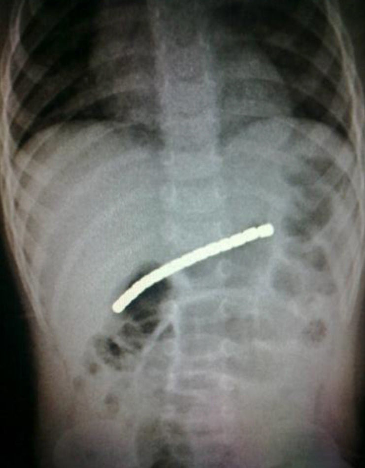
Clinical caseA previously healthy 23-month-old male child presented with colicky abdominal pain and hyporexia accompanied with vomiting of the gastrointestinal content. He was taken to a private-sector medical clinic where a plain abdominal x-ray was taken, revealing the presence of a radio-opaque object in the gastric chamber. He presented with an increase in abdominal pain and gastric content vomiting, for which he was referred to our institution. The parents stated in the interview that there had been no prior foreign object ingestion. The physical examination revealed a soft, depressible abdomen that was painful upon palpation at the epigastrium level; there were no signs of peritoneal irritation or acute abdomen. A plain abdominal x-ray was taken (6h after the first one) (fig. 1) and showed an approximately 9cm-long foreign body with a metallic aspect in the gastric chamber. The decision was made to perform an endoscopic study, in virtue of the symptomatology and the radiographic evidence.
.")
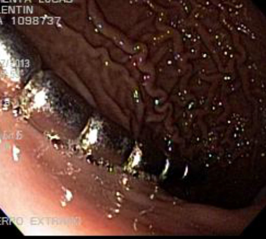
Diagnostic and therapeutic video panendoscopy was carried out 19h from the onset of the clinical symptoms; it identified a 10cm-long metal foreign body made up of 15 oval-shaped pieces measuring 4mm in width and 5mm in length, that were adhered to one another, and situated at the greater curvature of the stomach. The foreign body was extracted with a forceps and through laryngoscopy with a Magill forceps (fig. 2). The gastric mucosa presented with some superficial erosions and erythema.
Discussion
The ingestion of a single, isolated magnet is innocuous, similar to that of other foreign bodies, but the presence of numerous magnets represents a greater risk. In the cases in which magnets are located in different bowel segments, the pressure on them could cause mucosal lesions: erosions, ulcers, ischemia, and necrosis, as well as lesions in the intestinal wall situated between them: perforation, peritonitis, bowel obstruction, and fistulas. The formation of small bowel volvules7 and intraperitoneal bleeding8 have been reported that merited wide bowel resection, leading to short bowel syndrome and greater mortality.
Plain abdominal radiography can be useful for establishing the diagnosis. In the cases of ingestion of multiple magnets, they can be drawn together or lined up and give the impression that they are in the same place: «a single object». The lack of movement in the control x-rays can be secondary to being trapped in a bowel segment and producing the complications described above.8–10
The absence of clinical manifestations should not exclude aggressive intervention in cases of multiple ingested magnets located in different segments of the gastrointestinal tract.
An algorithm for pediatric population management by Hussain et al. (NASPGHAN 2012) has recently been published:
- 1.
Make diagnosis with the presence of gastrointestinal symptoms and/or small magnet antecedents; plain abdominal x-ray.
- 2.
Determine whether they are single or multiple and if there are metal objects, through x-rays in different positions; the latter 2 cases should be treated as emergencies due to the high risk for perforation.
- 3.
X-rays every 8-12h are recommended if the foreign body is in the intestine in order to evaluate its progression; if it has not moved in 24h, endoscopic or surgical removal of the object is indicated. No cases of spontaneous elimination of multiple magnets have been reported.4
Suspicion and awareness of the complications associated with the accidental ingestion of single or multiple magnets, with or without accompanying metal objects, are essential on the part of health care providers and family members so that early and opportune diagnosis and treatment can be carried out. In the present case of multiple magnets that were arranged in a line in the gastric chamber, the magnets did not progress into other segments of the gastrointestinal tract, enabling endoscopic management with no complications.
Please cite this article as: Cadena-León JF, Cázares-Méndez M, Arguello-Bermeo C, Cervantes Bustamante R, Ramírez-Mayans JA. Ingestión accidental de imanes en Pediatría: un problema de salud emergente. Revista de Gastroenterología de México. 2015;80:113–115.

