
A 67-year-old man with an unremarkable past medical history was receiving medical attention for 2 months due to odynophagia and progressive, high dysphagia to solid foods, as well as an approximate weight loss of 7kg.
The patient did not complain of respiratory symptomatology and stated he was not a smoker.
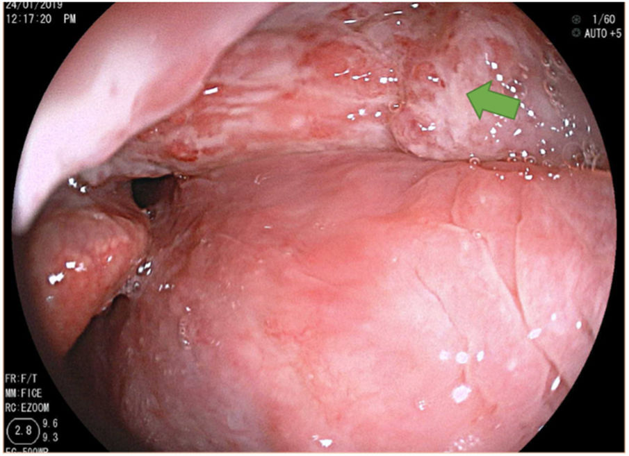
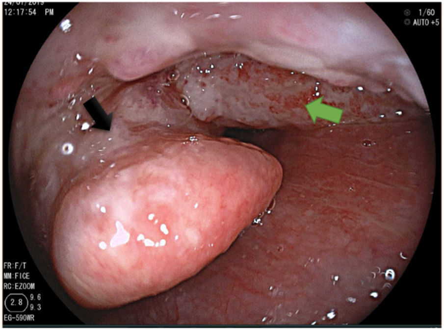
He was previously evaluated at the otorhinolaryngology service, and because a fiber optic laryngoscope was not available, evaluation at the gastroenterology service was requested. A barium esophagogram was performed and its results were normal. Considering that some segments of the respiratory tract are visualized during upper gastrointestinal endoscopy, the procedure was carried out. No lesions were found at the level of the esophagus, stomach, or duodenum, but during the removal of the equipment, the hypopharynx was examined, identifying a friable, granulomatous lesion located in the right glossoepiglottic fold extending to the epiglottis (Figs. 1 and 2). Multiple biopsy samples were taken. The histochemical Ziehl-Neelsen stain was positive, indicating tuberculosis. The patient was HIV negative and a chest tomography scan showed no lesions, confirming the diagnosis of primary hypopharyngeal tuberculosis. Anti-tuberculosis drug therapy was begun, and the patient’s progression is currently satisfactory.
.")
, extending to the epiglottis (black arrow).")
The authors declare that no experiments were performed on humans or animals for this study. No patient data appear in this article, guaranteeing confidentiality and anonymity to all participating patients.
Financial disclosureNo specific grants were received from public sector agencies, the business sector, or non-profit organizations in relation to this study.
Conflict of interestThe authors declare that there is no conflict of interest.
Please cite this article as: Mayorga-Garcés A, Hernandez S, Otero-Regino W. Alcance diagnóstico de la endoscopia digestiva: tuberculosis hipofaríngea. Revista de Gastroenterología de México. 2020;85:356–357.

